

Product overview
| Name | DAPI |
| Biological description | Overview DAPI is a blue fluorescent DNA stain which is cell permeant at high concentrations. DAPI binds strongly to A-T rich regions in DNA to form a fluorescent complex. It preferentially stains ds-DNA and has a high quantum yield (φf=0.92) when bound to DNA. Uses and applications DAPI is commonly used as a nuclear and chromosome counterstain. It is preferentially used to stain dead cells. DAPI is less effective as a live cell stain as it is unable to efficiently pass through the membrane in live cells. Therefore, higher concentrations may need to be used. Cells must be permeabilized and/or fixed for DAPI to enter the cell and bind to DNA. Due to DAPI’s blue emission, there is very little fluorescent overlap between yellow-fluorescent, green-florescent molecules (e,g, fluorescein and GFP) or red-fluorescent stains (e.g. Texas red). It is therefore convenient for multiplexing assays. DAPI has a great variety of applications but is often used for cell imaging, cell counting, cell sorting (based on DNA content), apoptosis analysis and in HCA (high-content analysis).
DAPI Staining Solution (1mg/mL) also available. |
| Purity | >98% |
| Description | Blue fluorescent DNA stain. Nuclear counterstain. Also available in solution. |
Images

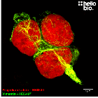
Figure 1. Neurofilament L and DAPI co-staining in hippocampal cell culture.

Figure 2. GFAP and DAPI co-staining in hippocampal cell culture

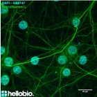
Figure 3. MAP2 and DAPI co-staining in hippocampal cell culture

Figure 1. Neurofilament L and DAPI co-staining in hippocampal cell culture.
Figure 2. GFAP and DAPI co-staining in hippocampal cell culture
Figure 3. MAP2 and DAPI co-staining in hippocampal cell culture
DAPI product vial image | Hello Bio
Biological Data
| Application notes | Figure 1: Neurofilament L and DAPI co-staining in hippocampal cell culture. DAPI is a DNA binding dye commonly used to label cell nuclei in immunofluorescence experiments. DAPI from Hello Bio labels cell nuclei (blue) at 1µg/ml when co-stained with an anti-neurofilament L antibody (green). For protocol see #Protocol 1 in application notes below.
Figure 2: GFAP and DAPI co-staining in hippocampal cell culture. DAPI is a DNA binding dye commonly used to label cell nuclei in immunofluorescence experiments. DAPI from Hello Bio labels cell nuclei (blue) at 1µg/ml when co-stained with an anti-GFAP antibody (green). For protocol see #Protocol 1 in application notes below.
Figure 3: MAP2 and DAPI co-staining in hippocampal cell culture. DAPI is a DNA binding dye commonly used to label cell nuclei in immunofluorescence experiments. DAPI from Hello Bio labels cell nuclei (blue) at 1µg/ml when co-stained with an anti-MAP2 antibody (green). For protocol see #Protocol 1 in application notes below.
#Protocol 1: DAPI counterstaining of primary cultured neurones.
|
Solubility & Handling
| Storage instructions | -20°C |
| Solubility overview | Soluble in water (10mg/ml, gentle warming), and in methanol |
| Storage of solutions | Prepare and use solutions on the same day if possible. Store solutions at -20°C for up to one month if storage is required. Equilibrate to RT and ensure the solution is precipitate free before use. |
| Shipping Conditions | Stable for ambient temperature shipping. Follow storage instructions on receipt. |
| Important | This product is for RESEARCH USE ONLY and is not intended for therapeutic or diagnostic use. Not for human or veterinary use. |
Chemical Data
| Purity | >98% |
| Chemical name | 4',6-Diamidino-2-phenylindole dihydrochloride |
| Molecular Weight | 350.24 |
| Chemical structure | ![DAPI [28718-90-3] Chemical Structure](https://cdn.hellobio.com/media/catalog/product//h/b/hb0747.png "DAPI [28718-90-3]") |
| Molecular Formula | C16H15N5.2HCl |
| CAS Number | 28718-90-3 |
| PubChem identifier | 160166 |
| SMILES | C1=CC(=CC=C1C2=CC3=C(N2)C=C(C=C3)C(=N)N)C(=N)N.Cl.Cl |
| InChiKey | FPNZBYLXNYPRLR-UHFFFAOYSA-N |
| MDL number | MFCD00012681 |
| Excitation | 340 / 360nM (for ds-DNA) |
| Emission | 488 / 460nM (for ds-DNA) |
Technical guides
 Understanding purity and quality - a guide for life scientists
References for DAPI
-
The use of DAPI fluorescence lifetime imaging for investigating chromatin condensation in human chromosomes.
Estandarte et al (2016) Sci Rep. 16 : 6-31417 -
Analysis of Apoptosis and Necroptosis by Fluorescence-Activated Cell Sorting.
Wallberg et al (2016) Cold Spring Harb Protoc 4 : 087387 -
New insights into the in situ microscopic visualization and quantification of inorganic polyphosphate stores by 4',6-diamidino-2-phenylindole (DAPI)-staining.
Gomes FM et al (2013) Eur J Histochem 57(4) : e34. -
Labeling nuclear DNA using DAPI.
Chazotte B (2011) Cold Spring Harb Protoc 2011(1) : pdb.prot5556. -
Labeling nuclear DNA using DAPI.
Chazotte et al (2011) Cold Spring Harb Protoc 2011(1) : 5556
-
No evidence from complementary data sources of a direct glutamatergic projection from the mouse anterior cingulate area to the hippocampal formation.
Andrianova L et al (2023) eLife 12PubMedID: 37545394 -
No evidence from complementary data sources of a direct projection from the mouse anterior cingulate cortex to the hippocampal formation
Craig et al (2022) Biorxiv : https://doi.org/10.1101/2022.01.25.477805
Related Products
- Code:
- HB8199
Blue fluorescent DNA stain. Nuclear counterstain. 1mg/mL staining solution in water. Solid also available in 10mg and 50mg packs.