Cell imaging tools - low cost, high quality reagents & probes

- DNA fluorescent stains
- Fluorescent Ion indicators & ionophores
- Fluorescent GPCR ligands (Cellaura)
- Other fluorescent probes
- Labels, stains and dyes
DNA Fluorescent stains
Our range of low-cost DNA fluorescent stains includes:
DAPI is a blue fluorescent DNA stain which is cell permeant at high concentrations It binds strongly to A-T rich regions in DNA to form a fluorescent complex. It preferentially stains ds-DNA and has a high quantum yield (φf=0.92) when bound to DNA. DAPI is commonly used as a nuclear and chromosome counterstain. It is commonly used to stain dead cells as it is less effective as a live cell stain as it is unable to efficiently pass through the membrane in live cells. Therefore, higher concentrations may need to be used. Cells must be permeabilized and/or fixed for DAPI to enter the cell and bind to DNA. DAPI has a great variety of applications but is often used for cell imaging, cell counting, cell sorting (based on DNA content), apoptosis analysis and in HCA (high-content analysis).
Hoechst 33342 is a blue fluorescent DNA stain that is commonly used in fluorescent microscopy. It is frequently used as a nuclear stain to stain nuclei. It is excited by UV light. Hoechst 33342 is cell permeable and has greater cell permeability than Hoechst 33258. The stain can be used on both live and fixed cells and is often used as an alternative to DAPI. Hoechst 33342 binds to the AT-rich regions of the minor grove in DNA which renders it specific for nuclear chromatin. Its fluorescent intensity depends on the DNA content, chromatin structure and the position of the cell within the cell cycle.
Hoechst 33258 is Blue fluorescent DNA stain that is commonly used in immunofluorescent work. It is frequently used as a nuclear stain to stain nuclei. Excited by UV light, it is less cell permeable but slightly more water soluble than the similar DNA stain Hoechst 33342. Unlike Hoechst 33342, Hoechst 33258 is not an apoptotic inducer. The stain is used as substitute to DAPI and can be used on both live and fixed cells.
Propidium iodide is a widely used red-fluorescent intercalating agent that binds and labels nucleic acids. It is membrane impermeant and is therefore frequently used to selectively identify dead cells and is commonly used in flow cytometry to evaluate cell viability. It is often used in flow cytometry, fluorescent microscopy and confocal laser scanning microscopy applications.
View all DNA Fluorescent stainsFluorescent ion indicators & ionophores
Our range of low-cost fluorescent ion indicators & ionophores include:Calcium indicators & chelators
- Fura-2 is a widely used UV light-excitable, ratiometric calcium indicator, suitable for ratio-imaging microscopy. It exhibits excitation ratiometry at 340 nm/380 nm with emission at 505 nm.
- Fura-2 AM is cell permeant and can be loaded into live cells noninvasively by incubation. It's widely used for ratio-imaging microscopy and measuring intracellular calcium elevations in neurons and other excitable cells.
- BAPTA AM is a non-fluorescent, cell permeable Ca2+ chelator, useful for manipulation of cellular Ca2+ levels.
Calcium ionophores
- A23187 (Calcimycin) is a non-selective Ca2+ ionophore, and has an intrinsic fluorescence excitable by UV light. This makes it less useful with UV-excitable Ca2+ indicators, (eg.Fura-2) but it is still useful for long-wavelength Ca2+ indicators, e.g., Fluo-2.
- Ionomycin and ionomycin calcium salt are effective Ca2+ ionophores commonly used to both calibrate fluorescent Ca2+ indicators and to modify intracellular Ca2+ concentrations.
Fluorescent GPCR ligands - the CellAura range

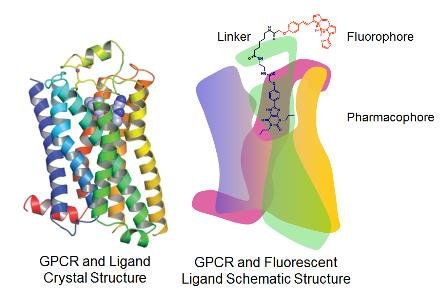
CellAura fluorescent ligands target G protein coupled receptors (GPCRs) and are comprised of three units:
- a pharmacophore - e.g. a synthetic agonist or antagonist
- a fluorescent dye (the fluorophore)
- a linker - which connects the pharmacophore with the dye
The CellAura range currently includes ligands that target GPCRs such as adenosine, dopamine, 5-HT, adrenergic, muscarinic and histamine receptors. Each CellAura fluorescent ligand has been characterised using live cell imaging and functional analysis to confirm its affinity and pharmacological activity. They have been used successfully in a wide range of applications including: fluorescent ligand binding, high content screening, live cell imaging of receptor-ligand binding,displacement and receptor internalisation, FCS, FACS, real-time analysis of single molecule ligand-receptor interactions, confocal microscopy, High Throughput Screening, and more.
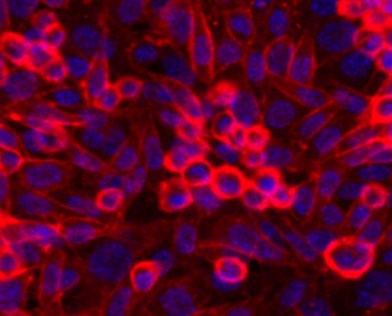
![Binding of CA200623 CellAura fluorescent adenosine agonist [NECA] Binding of CA200623 CellAura fluorescent adenosine agonist [NECA]](https://hellobio.com/media/wysiwyg/Products/HB7813-CA200623-image1-fl_result.jpg)
CA200634 CellAura fluorescent adenosine antagonist [XAC] is a competitive fluorescent adenosine receptor antagonist (with apparent KD values of 7.50, 7.37 and 7.30 for A2A, A3 and A1 respectively). It antagonizes the activity of NECA, an adenosine receptor agonist and exhibits no intrinsic agonist activity.
CA200623 CellAura fluorescent adenosine agonist [NECA] is a fluorescent adenosine receptor agonist (pEC50 values are 8.57, 8.47, 6.76 and 5.69 for A3, A1, A2A and A2B respectively), derived from the non-selective adenosine agonist NECA.
CA200843 CellAura fluorescent H3 antagonist [clobenpropit] is a fluorescent H3 histamine receptor antagonist (apparent KD values are 7.09, 6.55 and 5.71 for H3, H1 and H2 receptors respectively). It antagonizes the activity of histamine and displays no intrinsic activity.
Other Fluorescent probes
- Sulforhodamine 101 (SR101) is a red fluorescent dye which is water-soluble and non-fixable. It is a preferential astrocyte marker both in vitro and in vivo and is frequently used in neurophysiological experiments. It also labels oligodendrocytes.
- Calcein-AM is a non- fluorescent and cell permeable dye. Hydrolysed into green fluorescent calcein by cytosolic esterases in living cells its uses include monitoring chemotaxis, multidrug resistance and determining cell viability.
- View the full range
Labels, stains & dyes
BrdU (5-Bromo-2′-deoxyuridine) is a thymidine analog which is incorporated into DNA during DNA replication (during S-phase of cell cycle). It is widely used to identify proliferating cells and labels cell lines and primary cell cultures in vitro and also cells in vivo. View the full range of labels, stains and dyes.


